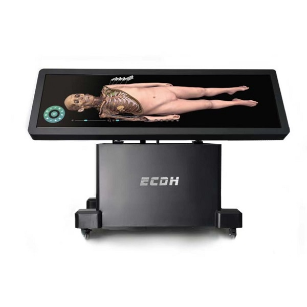
Virtual human dissection is transforming the way anatomy is taught and studied. Using digital technology, it replicates traditional dissection with highly detailed, 3D models of the human body. Unlike physical dissection, this method is safer, more efficient, and environmentally friendly. Virtual human dissection also allows for repeated access to anatomical structures, offering a more flexible learning experience than ever before.
Next-Level Anatomy Learning
The evolution of medical education is being driven by advanced technologies such as Virtual human dissection. Leading this innovation is the DIGIHUMAN Virtual Anatomy Table, a state-of-the-art tool that reconstructs the human body in ultra-high-definition 3D. Through intuitive multi-touch control, users can perform digital dissections, rotate anatomical structures, and explore internal systems layer by layer. This immersive experience enables students and professionals to gain a deeper understanding of both normal and pathological anatomy without the limitations of physical specimens. With highly realistic visuals and precise modeling, DIGIHUMAN offers a safe, repeatable, and effective way to master human anatomy.
Digital Tools for Deeper Insight
The DIGIHUMAN system features five integrated modules: Human Anatomy, Digital Human Anatomy System, Slice Library, Clinical Cases, and Embryology. This comprehensive setup provides realistic visualization of both normal and pathological anatomy. The interactive interface enhances learning by allowing detailed, repeatable exploration of anatomical systems. As a high-performance educational tool, Virtual human dissection using this system helps bridge the gap between theoretical knowledge and clinical application, making it an ideal solution for institutions embracing the future of medical training.
Conclusion
Virtual human dissection, especially with DIGIHUMAN, is the future of anatomy education, offering an advanced, interactive, and accessible solution for students and educators alike.

